รวมกัน ภาพถ่ายเกี่ยวกับภาพ ct scan ที่เว็บไซต์ littlestarcenter.edu.vn รวบรวมและจัดทำอย่างครบถ้วนค่ะ มีภาพถ่ายที่เกี่ยวข้องกับ ที่คุณสามารถดูรายละเอียดเพิ่มเติมที่ด้านล่างค่ะ
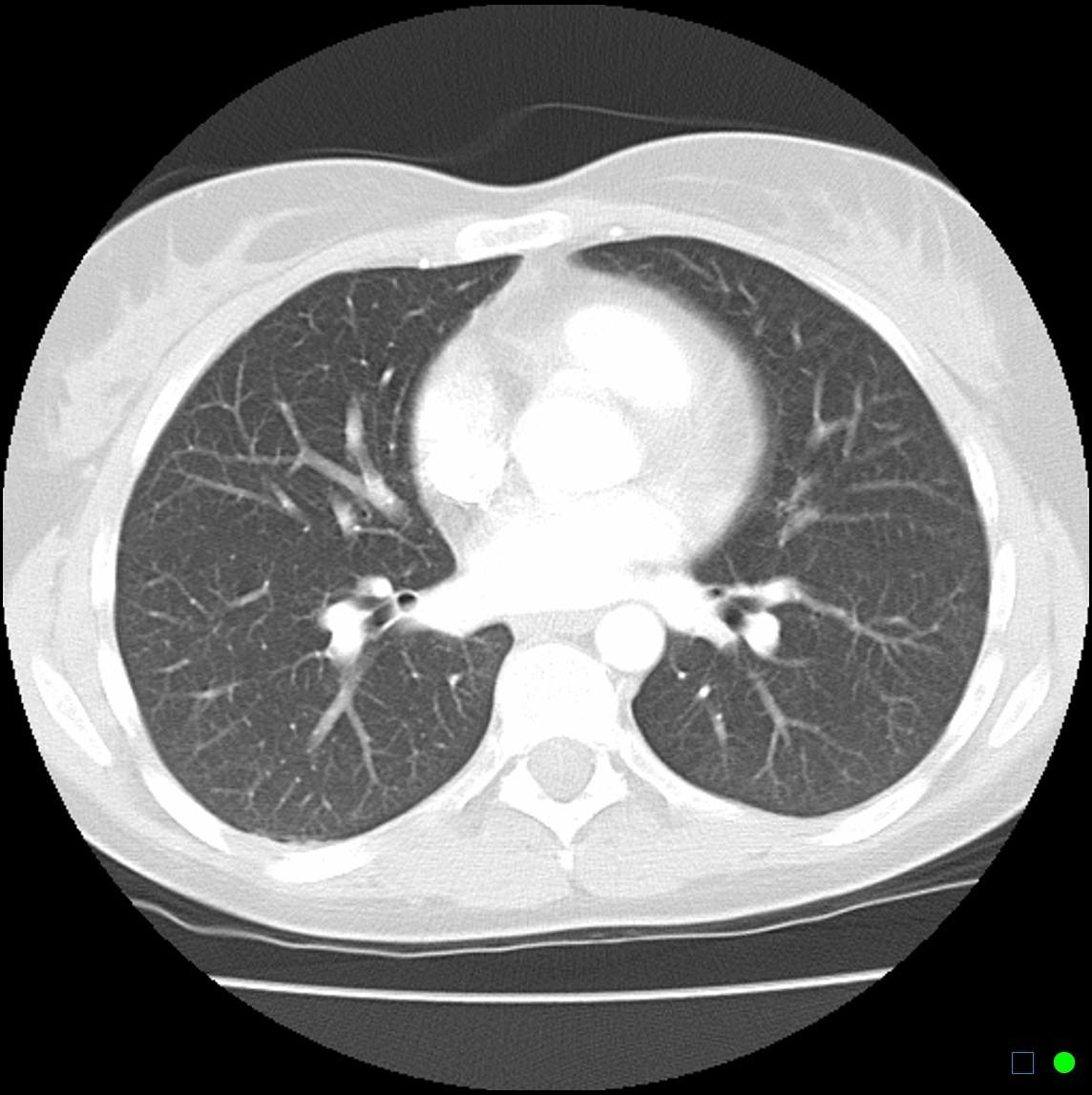
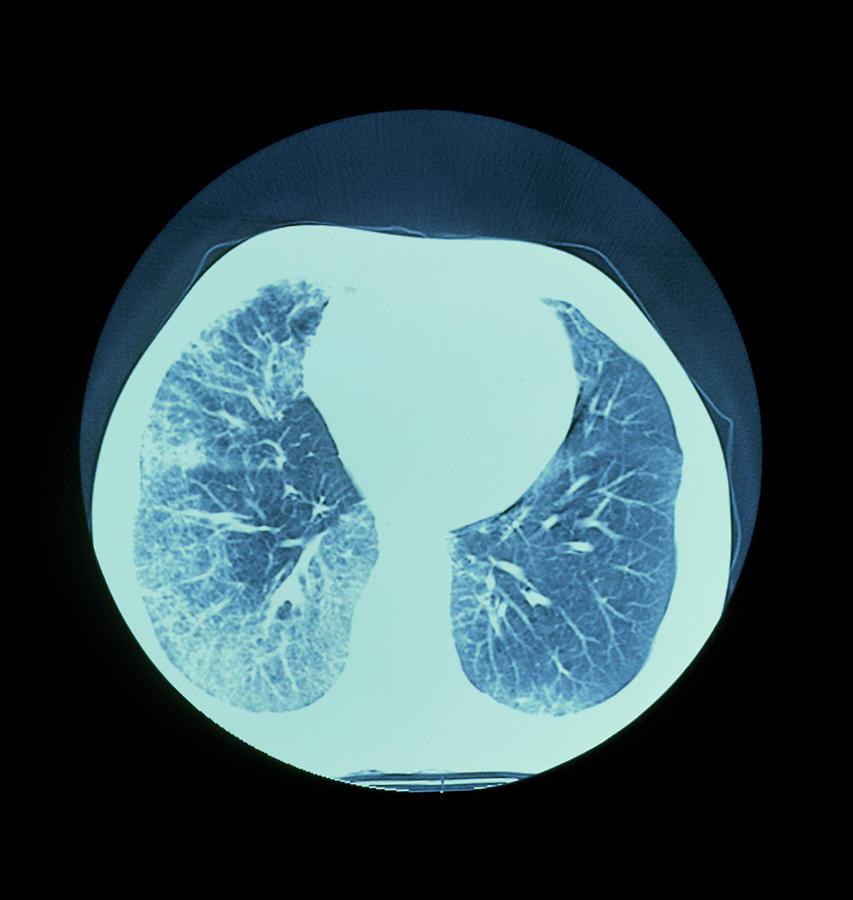
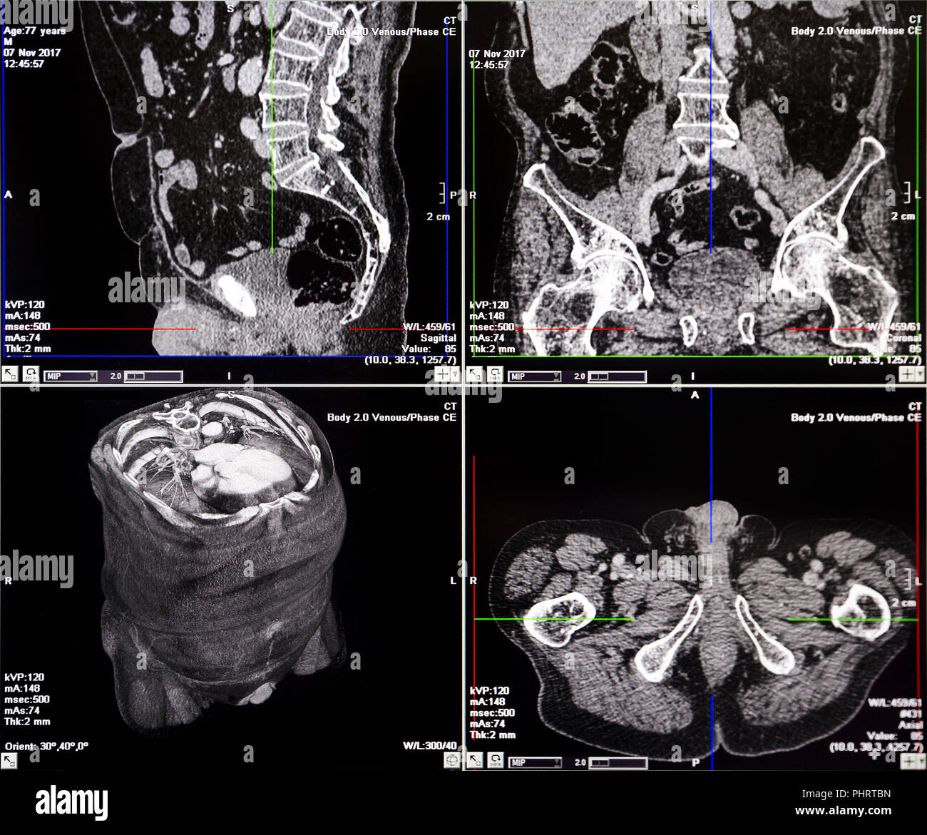
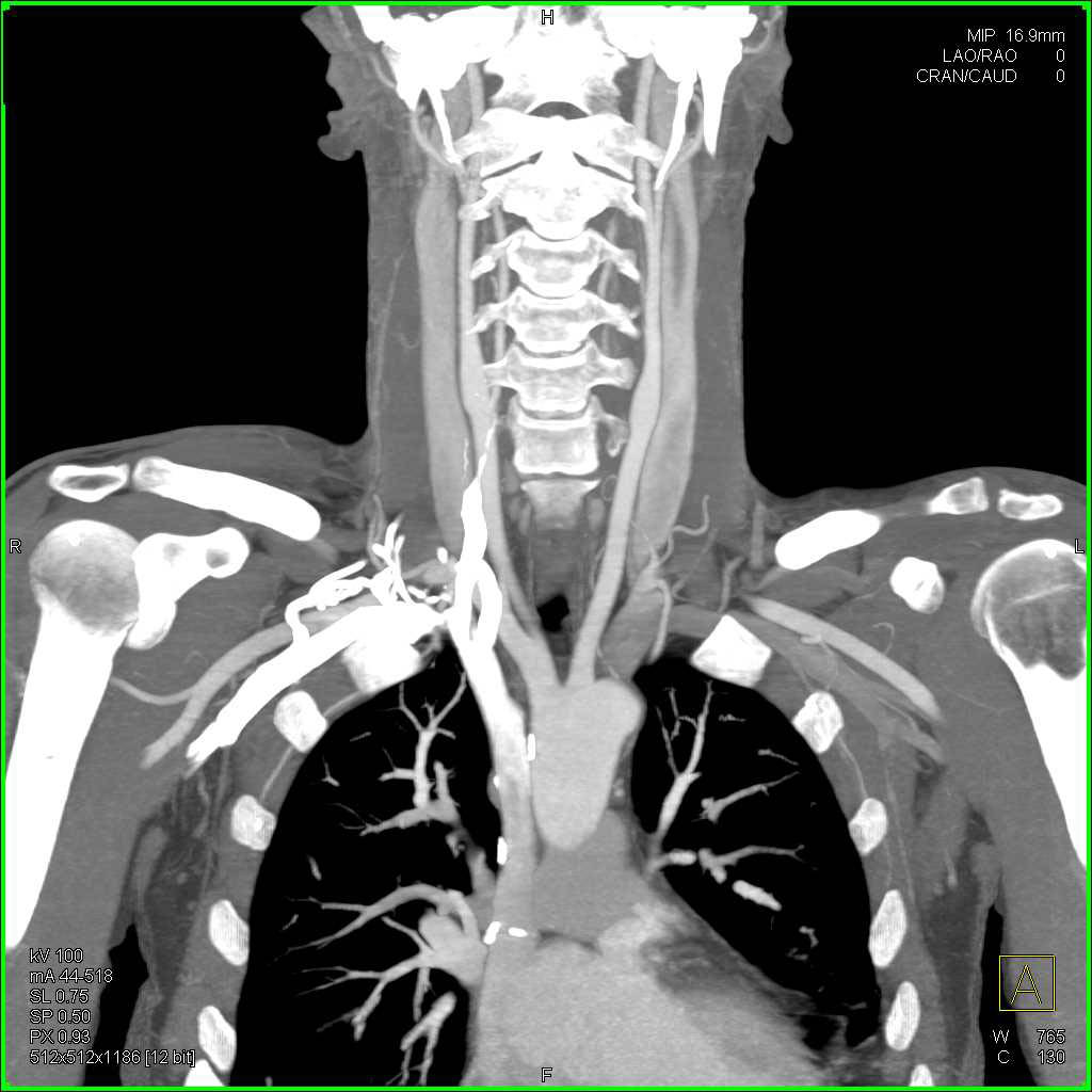
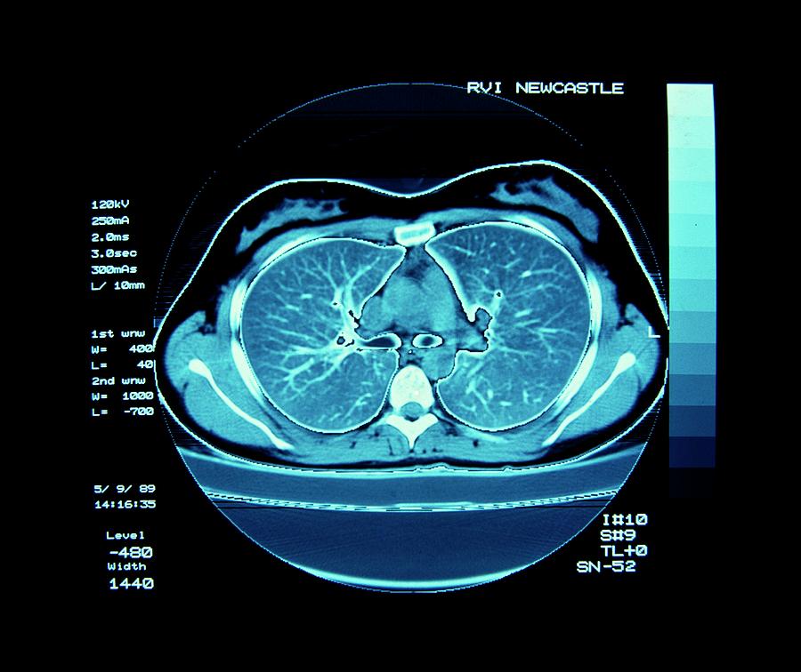
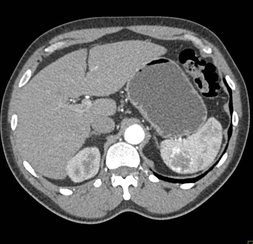
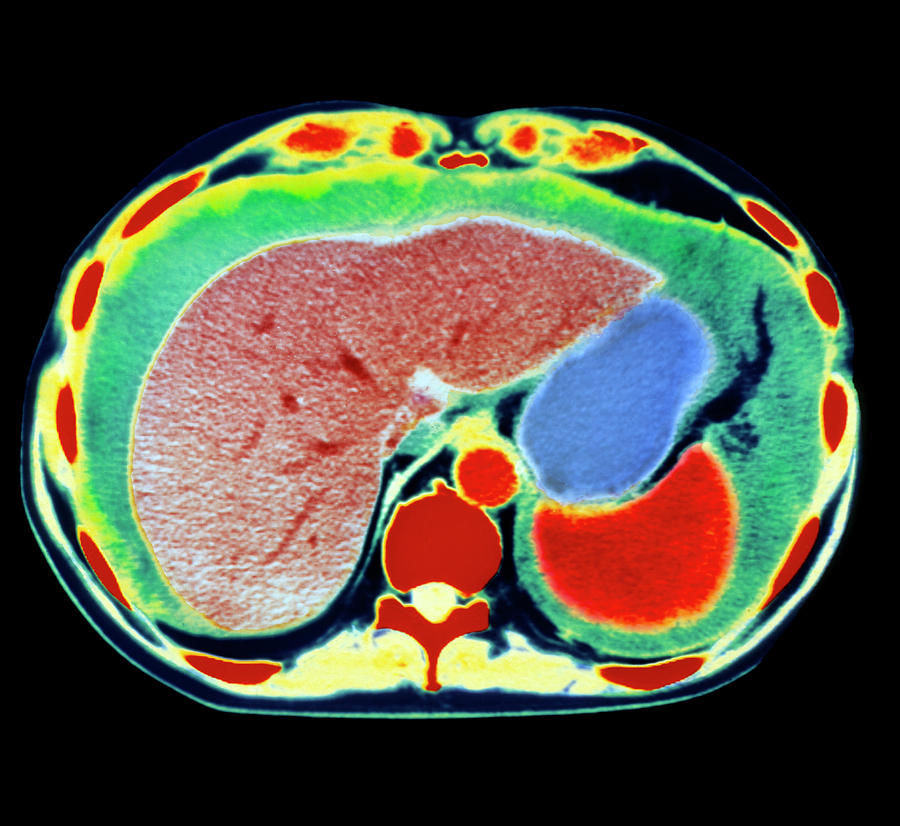
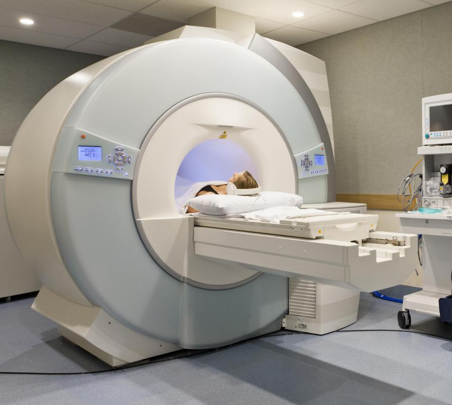
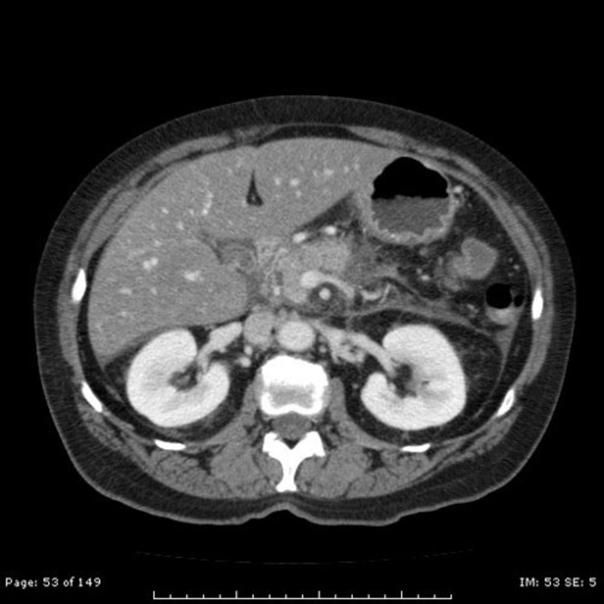
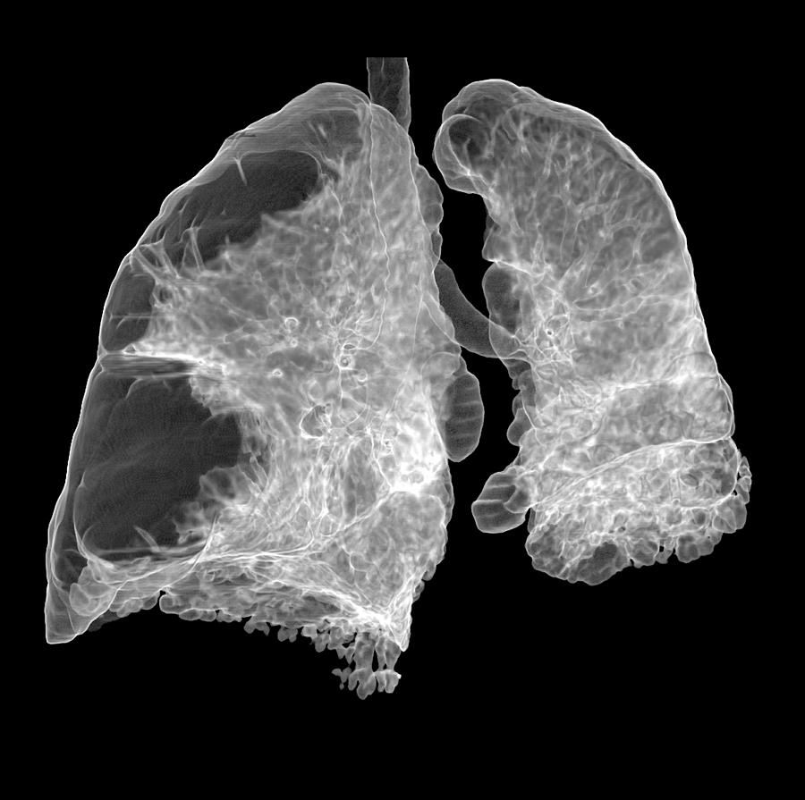
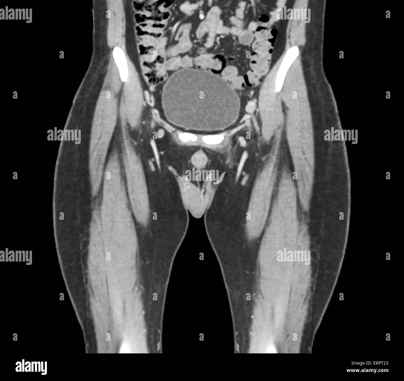
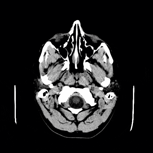
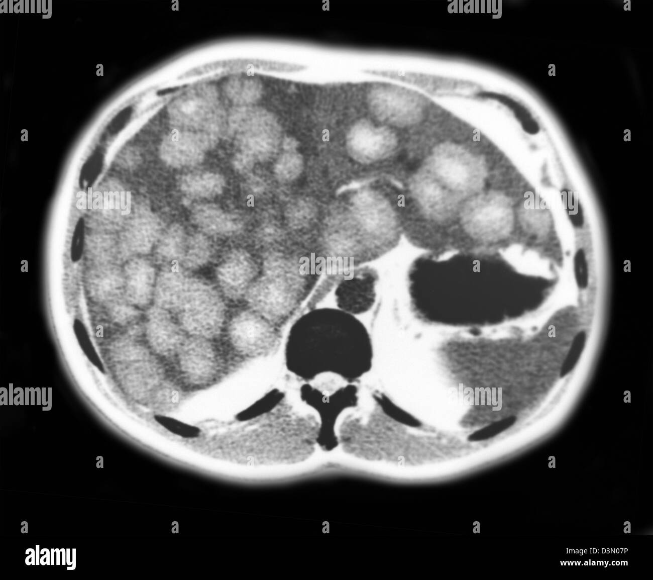
ภาพ ct scan


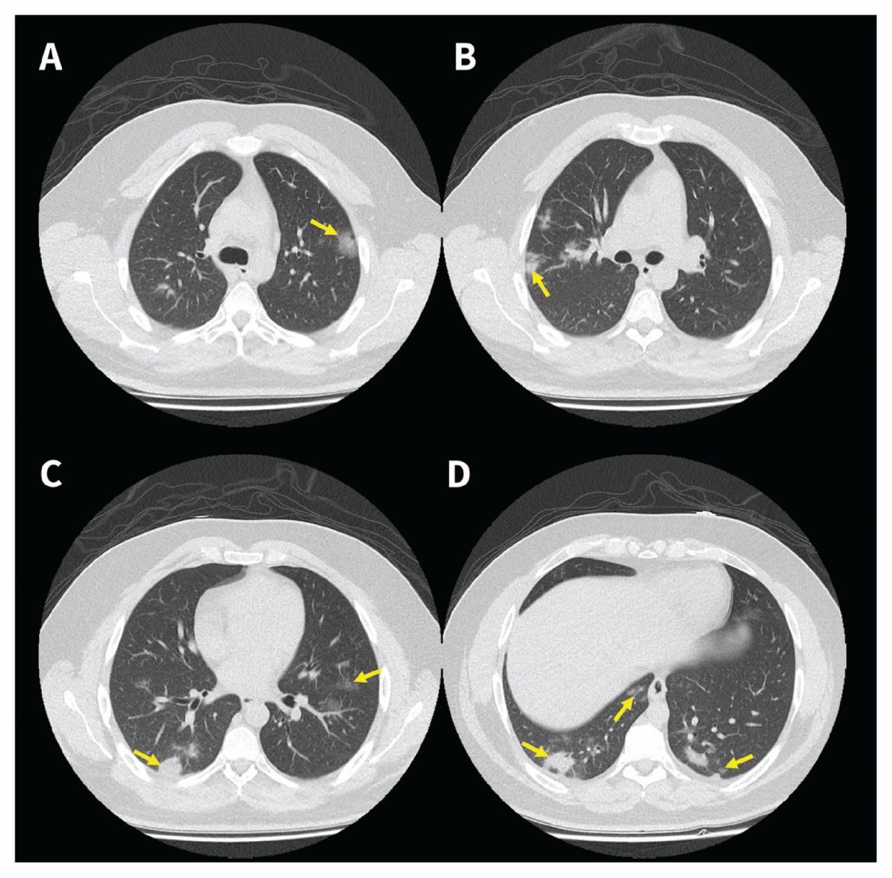


MRI or CT-scan report me kya batya doctor ne 🥺(ishugangore vlog)
ขอขอบคุณที่ให้ความสนใจในบทความ ภาพ ct scan ที่มีอยู่ที่ littlestarcenter.edu.vn ค่ะ คุณมีสิทธิ์ในการแสดงความคิดเห็น และยังสามารถตรวจสอบบทความที่เกี่ยวข้องเพิ่มเติมได้ที่ด้านล่างค่ะ หวังว่าจะช่วยเสริมสร้างข้อมูลที่น่าสนใจให้กับคุณค่ะ
Posts: ภาพ ct scan
Categories: อัลบั้ม
Author: littlestarcenter.edu.vn